Seminar: The Last Nanometer – The way water meets DNA and Solid Surfaces in X10,000,000 magnification

22/05/2019 - 12:00 - 11:00Add To Calendar 2019-05-22 11:00:00 2019-05-22 12:00:00 Seminar: The Last Nanometer – The way water meets DNA and Solid Surfaces in X10,000,000 magnification S E M I N A R Wednesday 22/05/19, 11:00 am Building 211, seminar room   SPEAKER: Prof. Uri Sivan Department of Physics, Technion-Israel Institute of Technology   TOPIC: The Last Nanometer – The way water meets DNA and Solid Surfaces in X10,000,000 magnification   Recent advancements in atomic force microscopy facilitate atomic-resolution three-dimensional mapping of hydration layers next to macromolecules and solid surfaces. These maps provide unprecedented information on the way water molecules organize around these objects and interact with them. After a short presentation of our home-built microscope, characterized by sub 0.1 Å noise level, I will focus on three representative studies. The first will disclose that water molecules grow epitaxially on certain hydrophilic crystalline substrates, forming 3d ice extending to about 1 nm from the surface. The second study will present our recent success in obtaining ultra-high-resolution images of DNA and 3d maps of its hydration structure (e.g., figure below). This study shows that labile water molecules concentrate along the DNA grooves, in agreement with known position of DNA binding sites. The third study will disclose our recent discovery that hydrophobic surfaces in contact with non-degassed water are generically coated with a layer of condensed gas molecules of a density about half that of liquid nitrogen. This layer not only renders the hydrophobic interaction a certain universality, but also identifies the source of hydrophobic attraction – one of the oldest puzzles of physical chemistry.         (a) An ultra-high-resolution image of DNA with a reference model of B-DNA. The major grooves, minor grooves and top-facing phosphates are highlighted with gray and white arrows on the model and the scan. Scale bar, 5nm. (b) Hydration of double stranded DNA. Red shaded pixels mark the position of labile water molecules. Abstract Department of Chemistry chemistry.office@biu.ac.il Asia/Jerusalem public

S E M I N A R

Wednesday 22/05/19, 11:00 am

Building 211, seminar room


SPEAKER:

Prof. Uri Sivan

Department of Physics,

Technion-Israel Institute of Technology


TOPIC:

The Last Nanometer – The way water meets DNA and Solid Surfaces in X10,000,000 magnification


Recent advancements in atomic force microscopy facilitate atomic-resolution three-dimensional mapping of hydration layers next to macromolecules and solid surfaces. These maps provide unprecedented information on the way water molecules organize around these objects and interact with them.

After a short presentation of our home-built microscope, characterized by sub 0.1 Å noise level, I will focus on three representative studies. The first will disclose that water molecules grow epitaxially on certain hydrophilic crystalline substrates, forming 3d ice extending to about 1 nm from the surface. The second study will present our recent success in obtaining ultra-high-resolution images of DNA and 3d maps of its hydration structure (e.g., figure below). This study shows that labile water molecules concentrate along the DNA grooves, in agreement with known position of DNA binding sites. The third study will disclose our recent discovery that hydrophobic surfaces in contact with non-degassed water are generically coated with a layer of condensed gas molecules of a density about half that of liquid nitrogen. This layer not only renders the hydrophobic interaction a certain universality, but also identifies the source of hydrophobic attraction – one of the oldest puzzles of physical chemistry.
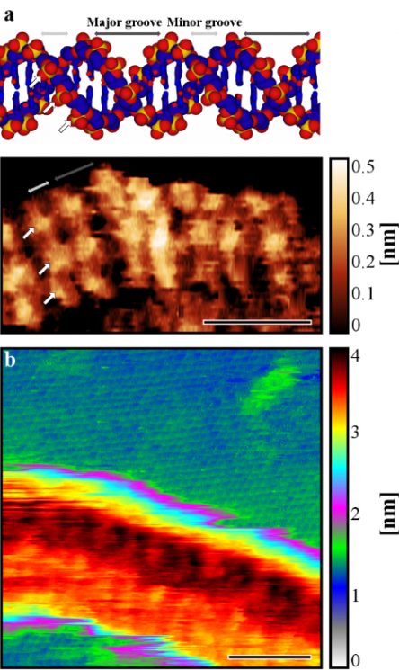
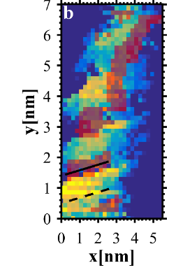


(a) An ultra-high-resolution image of DNA with a reference model of B-DNA. The major grooves, minor grooves and top-facing phosphates are highlighted with gray and white arrows on the model and the scan. Scale bar, 5nm. (b) Hydration of double stranded DNA. Red shaded pixels mark the position of labile water molecules.

Abstract

Last Updated Date : 20/05/2019